Sunday, November 23, 2008
Bullous Pemphigoid


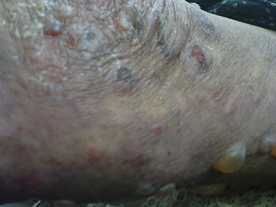
Bullous pemphigoid (BP) is a chronic blistering of the skin. It ranges from mildly itchy welts to severe blisters and infection, and may affect a small area of the body or be widespread. The vast majority of those affected are elderly, but it has been seen at all ages.
It is an autoimmune disorder, meaning it is caused when the body's immune system malfunctions. The immune system is meant to defend the body against bacteria, viruses, and disease, but instead produces antibodies against healthy tissue, cells and organs. Some patients with BP have other autoimmune diseases such diabetes and rheumatoid arthritis. Various other factors have been reported to play a role in triggering BP. These include drugs (furosemide, penicillin's), mechanical trauma, and physical traumas (burns from radiation, sun or heat).
Bullous is the medical term for a large blister (a thin-walled sac filled with clear fluid). Usually the skin in BP is very itchy and large, red welts and hives may appear before or during the formation of blisters. The blisters are widespread and usually appear on the areas of the body that flex or move (flexural areas). About 15-20 percent of people with BP also develop blisters in the mouth or down the throat in the esophagus.
Because of all the variations and differing degrees of symptoms, the diagnosis must be confirmed by skin biopsy. A special skin biopsy test (a direct immunoflorecsence biopsy) may also be needed. Blood tests are usually inconclusive.
Treatment is focused on relief of symptoms and prevention of infection. Tetracycline and Minocycline antibiotics are very useful for mild to moderate disease. They do not work on bacteria, but act directly on the immune system. They can be used in combination with potent topical steroid creams for more rapid relief.
Oral steroids (prednisone, prednisolone) are the treatment of choice for severe cases. Regular visits will be needed because the dose must be adjusted frequently, and side effects must be monitored. A fairly high dose is needed initially, and once the blisters have stopped appearing, it is slowly reduced over many months or years. As steroids have some undesirable side effects, dermatologists try to reduce the dose as low as possible. If this is done too quickly, the blisters reappear.
Often, immunosuppressive agents (Immuran, Cellcept, Methotrexate, cyclophosphamide and Neoral) are used in combination with the oral steroids to allow a lower dose. Severe cases are best treated in the hospital to allow expert dressing of the wounds, and intravenous injections of the most potent treatments.
BP is a self-limiting disease that is in most cases eventually completely clears up and the treatment can be stopped. Treatment is usually needed for several years, but generally after a few months it is possible to reduce the dose of medications to reasonably low levels. BP also often has a pattern of remissions and flare-ups. It may be without symptoms for 5 or 6 years then suddenly flare up.
With careful management, most patients with BP do well. Be patient and faithfully follow your instructions, these are the keys to good results
Labels: Bullous Pemphigoid, Dermatology
Leishmaniasis
we tested him for leishmaniasis for 3 times and all of them was negative, but at last montenegro skin test and skin biopsy became positive and cutaneous leishmaniasis confirmed
Differential diagnosis list was:
- Lupus vulgaris
- leishmaniasis
- Sarcoidosis


Labels: Dermatology, Leishmaniasis
Seborrheic keratosis

Definition
Seborrheic keratosis is one of the most common types of noncancerous (benign) skin growths in older adults. In fact, most people develop at least one seborrheic keratosis at some point in their lives.
A seborrheic keratosis usually appears as a brown, black or pale growth on the face, chest, shoulders and back. The growth has a waxy, scaly, slightly elevated appearance. Occasionally, it appears singly, but multiple growths are more common. Typically, seborrheic keratoses don't become cancerous, but they can look like skin cancer.
These skin growths are normally painless and require no treatment. You may decide, however, to have them removed if they become irritated by clothing or for cosmetic reasons.
Symptoms
A seborrheic keratosis usually has the appearance of a waxy or wart-like growth. It typically appears on the head, neck or trunk of the body. A seborrheic keratosis:- Ranges in color from light tan to black
- Is round- to oval-shaped
- Has a characteristic "pasted on" look
- Is flat or slightly elevated with a scaly surface
- Ranges in size from very small to more than 1 inch (2.5 centimeters) across
- May itch
It may develop a single growth or cluster of growths. Though not painful, seborrheic keratoses may prove bothersome depending on their size and location. Be careful not to rub, scratch or pick them. This can lead to inflammation, bleeding and infection.
CausesThe exact cause of seborrheic keratoses is unclear. They tend to run in some families, so genetics may play a role. Ultraviolet (UV) light may also play a role in their development since they are common on sun-exposed areas, such as the back, arms, face and neck.
Tests and diagnosis
Diagnose seborrheic keratosis made by inspecting the growth. To confirm the diagnosis or to rule out other skin conditions, biopsy may be necessary. Typically, seborrheic keratosis doesn't become cancerous, but it can resemble skin cancer.
Treatments and drugs
Treatment of seborrheic keratoses usually isn't necessary. However, patient may want them removed if they become irritated, if they bleed , or if the patient simply don't like how they look or feel.
This type of growth is never deeply rooted, so removal is usually simple and not likely to leave scars:
- Cryosurgery. Cryosurgery can be an effective way to remove seborrheic keratosis. However, it may not work on large, thick growths, and it may lighten the treated skin (hypopigmentation).
- Curettage. Sometimes curettage is used along with cryosurgery to treat thinner or flat growths. It may be used with electrocautery.
- Electrocautery. Used alone or with curettage, electrocautery can be effective in removing seborrheic keratosis. This procedure can leave scars if it's not done properly, and it may take longer than other removal methods.
Medical reasons for seborrheic keratosis treatment include intense itching, pain, inflammation, bleeding and infection.
Labels: Dermatology, Seborrheic Keratosis


